Inside an MRI Scanner: Component by Component
The design of an MRI scanner is a bit like the interior of a building, where each room has a different job. From the outside you mostly see the tube. Behind the casing, however, several components work at once. What do they look like when you go through the scanner from the inside out?
Receiver Coils and the Transmit Unit on the Inside
The radio system works closest to the body region being scanned. The transmit unit sends out pulses, while the receiver coils pick up the signal that comes back from that region. Because receiver coils are fitted to the body region in question, a scanner cannot get by with a single universal receiver. The signal that is recorded forms the basis of the image later on. The patient table also belongs to this inner area, as a fixed part of the whole system.
Gradient Coils: Where Does the Knocking Come From?
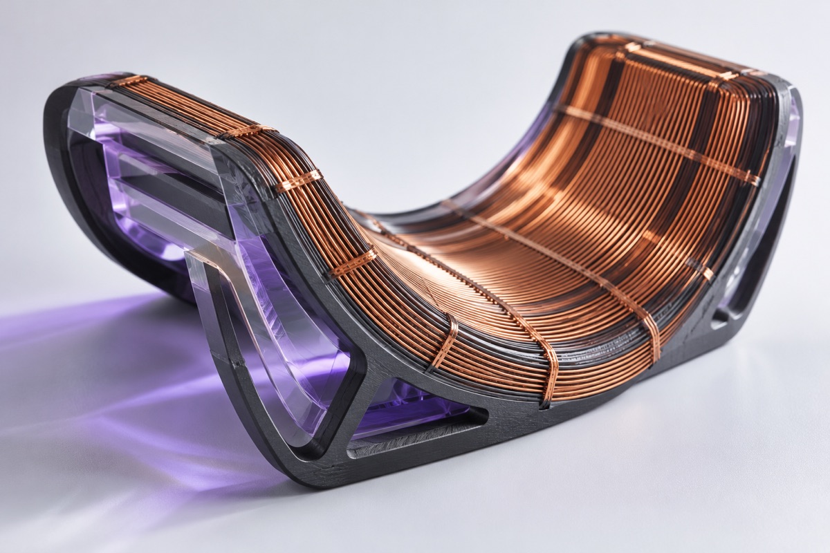
The gradient coils sit in the tube. They are cylindrical and lie inside the bore of the main magnet. There they produce fields that vary across space. Compared with the main field these fields are small, but they cause the characteristic knocking and hammering during a scan. The noise is therefore tied to how the gradient coils operate.
The Superconducting Magnet: The Heart of the Scanner
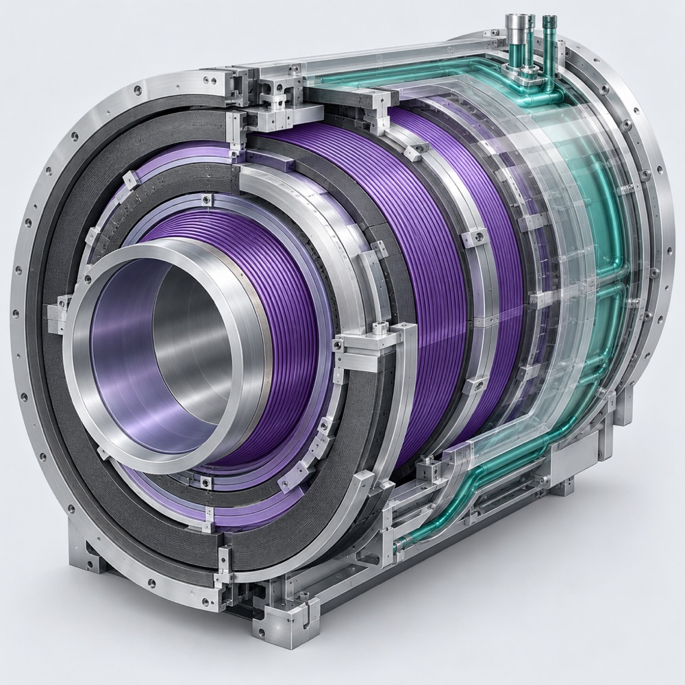
The central part of the whole construction is the magnet. It produces a strong, steady field known as the main field. Many practices and hospitals run scanners rated at 1.5 or 3 tesla. Many scanners use a superconducting magnet that is cooled to about 4 kelvin with liquid helium. At this very low temperature, the electrical resistance of the coil practically disappears.
The magnet sits inside a cooling vessel. This vessel consists of a vacuum housing, a heat shield and a tank for the liquid helium. The layers are arranged like the walls of a house, with each one protecting what lies further in.
Is Liquid Helium Always Required?
Not always, since scanners using high-temperature superconductors have also been developed, and they manage without liquid helium. For now this is a matter of technical development. When we describe the design of an MRI scanner here, we usually mean the classic superconducting magnet. Even so, it is worth keeping in mind that this part of the construction may change in the future.
Computer and Shielded Room: The Outer Layer
The scanner stands in a shielded room, which forms the last layer of the whole system. Next to it works the control computer. It converts the collected signals into cross-sectional images. This is how the raw data from the receiver coils becomes a picture you can look at.
Summary
If you know how an MRI scanner is built, you understand how its different parts work together: receiver coils and transmit unit, gradient coils, superconducting magnet, patient table, computer and shielded room. Each one has a job of its own. You will find more articles on the technology on the other pages of Spinschicht.