How Does an MRI Work? From Magnetic Field to Image
The working principle of magnetic resonance imaging comes down to this: coils pick up a radio signal from a body that was first placed in a strong magnetic field. Only after several steps does that signal become a cross-section you can view on a monitor. This article walks through how it happens, one step at a time.
The Magnetic Field Lines Up the Hydrogen Nuclei
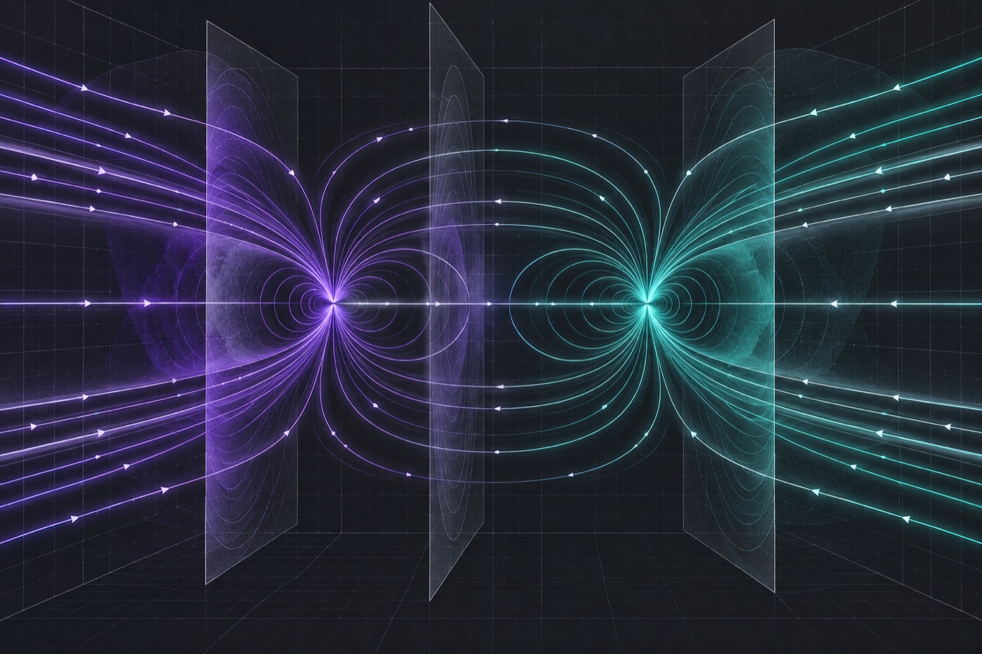
Everything starts with a strong, uniform magnetic field. The body holds a lot of water and fat, and both contain hydrogen nuclei. That is what makes tissue such a good source of signal for magnetic resonance: the nuclei are present in huge numbers and together give off a signal that can be received. Every nucleus has a property called spin, which makes it act like a tiny magnet.
Under the influence of the main field, the spins of the hydrogen nuclei in the tissue line up along that field. This creates the starting state for the whole measurement. One point matters here: the machine does not measure anything yet during this step. It only prepares the tissue for what comes next.
The Radio Pulse and the Signal From the Tissue
The second step is a radio pulse at a carefully chosen frequency. It excites the aligned hydrogen nuclei. The match between the two frequencies is exactly the resonance that gives the method its name. After the pulse, the nuclei return to their starting state and send out a radio signal as they do.
Coils in the scanner pick up this signal. They convert the very weak waves coming from the tissue into data that can be processed further. So magnetic resonance imaging does not shine anything through the body. It receives a signal that the tissue produces by itself.
How Gradients Pinpoint Location in the Body
The signal from the hydrogen nuclei alone does not reveal where it came from. The gradient coils supply the missing location information. They add a gentle slope in field strength, in three directions, on top of the main field. Nuclei at different positions therefore feel a slightly different field, so their signal can be assigned to a specific point in space.
Gradients are thus the key to localization. Without them, the machine would notice that the tissue is sending a signal but could not tell where it originates. Because the slope acts in three directions, position can be worked out across the whole slice under examination, not just along a single line. That makes this step just as important as exciting the nuclei.
The Computer Calculates the Cross-Section
The computer handles the final step. It calculates a cross-sectional image from the collected signals. The mathematics behind this relies on the Fourier transform. In practice, the raw data from the coils is translated into bright and dark areas that correspond to different tissues. Each image point belongs to the location that the gradients assigned earlier.
This is another way the principle differs from computed tomography. Unlike CT, magnetic resonance imaging works without ionizing radiation. The picture is created purely from a magnetic field and radio waves.
Summary
The working principle can be described in five steps in a row. The field lines up the hydrogen nuclei, the radio pulse excites them, coils receive the signal, gradients fix its location, and the computer assembles the data into an image. Once you understand this order, it is easier to see how the method differs from other imaging techniques. Feel free to read the other articles on Spinschicht as well. They are written as popular science and do not replace medical advice.